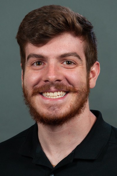
BME Team: (from L to R) Justin Hollenbeck, Adam Bradshaw, Justin Brown (RA), Mitch Mologne, Scott Tashman, Colin Smith, Austin Carcia, Alex Brady, Steve Atherton, Brad Fossum (RA), Lauren Watkins.
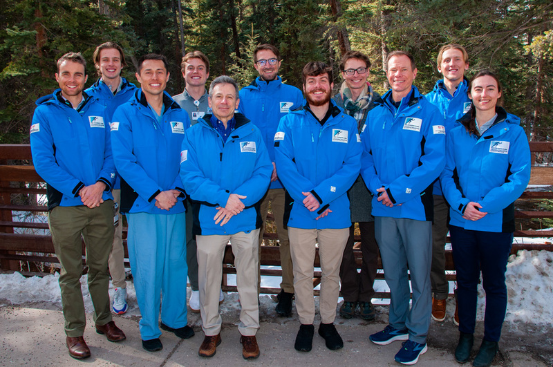
BME Team: (from L to R) Justin Hollenbeck, Adam Bradshaw, Justin Brown (RA), Mitch Mologne, Scott Tashman, Colin Smith, Austin Carcia, Alex Brady, Steve Atherton, Brad Fossum (RA), Lauren Watkins.
SPRI’s Department of Biomedical Engineering is a collection of multidisciplinary laboratories including Biomotion, Robotics and Advanced Imaging. The team is comprised of scientists and researchers that apply quantitative, analytical and integrative methods to the field of orthopaedic medicine.
In 2021, the Department of Imaging Research officially joined the Department of Biomedical Engineering (BME), further integrating the labs and research projects. The team employs three full-time PhD researchers in Director Dr. Scott Tashman and Research Scientists Drs. Lauren Watkins and Colin Smith.
The BME team focuses on the role of mechanics and movement on injury and re-injury prevention as well as assessment of surgical and nonsurgical treatments for restoring musculoskeletal function. The team is dedicated to integrating clinical care, research and education, combining SPRI’s engineering expertise with the resources of renowned orthopaedic surgeons from The Steadman Clinic, with the ultimate goal of improving treatment of musculoskeletal diseases and orthopaedic injuries. Biomechanics, motion analysis, advanced biomedical imaging, computational modeling and orthopaedic engineering are at the center of the department’s research approach.
BME continues to collaborate closely with physicians, maintaining the imperative clinical lens that keeps its focus on high-impact research. BME’s research portfolio has expanded significantly to include an essential role in nearly all of SPRI’s clinical trials, providing advanced biomechanical and quantitative imaging assessments to evaluate the effectiveness of regenerative medicine treatments for restoring tissue health and function.
The department is home to a state-of-the-art Biomotion Laboratory for assessing human movement and function that includes video-motion analysis, an instrumented treadmill, force plates, a wireless EMG system, wearable IMU sensors, insole pressure sensors and a unique Dynamic Stereo X-ray system, utilizing cutting-edge technology designed by Dr. Scott Tashman, BME Director. Studies of musculoskeletal anatomy and new orthopaedic procedures are conducted in the department’s Robotics Laboratory, which is one of the most advanced facilities of its kind in the world. This includes a custom robotic-arm that can recreate physiological joint motion, and video-motion analysis and 3D laser scanning technologies to capture musculoskeletal geometry and function. The BME team also conducts advanced imaging with its leading-edge 3.0 Tesla Siemens MAGNETOM Skyra and Canon Vantage Galan magnetic resonance imaging (MRI) scanners in Vail, Frisco and Basalt, Colorado, while applying imaging tools to improve patient outcomes.
The SPRI Golf Sports Medicine Program operates under the Department of Biomedical Engineering, integrating a high-tech simulator system within the Biomotion Laboratory. This unique combination of technologies enables the SPRI to perform one of the most advanced biomechanical analyses of golf swings in the world, leading to a faster return to play after orthopaedic treatments, reduced injuries, and improved performance.
Medical imaging plays a vital role in orthopaedic and sports medicine for diagnosis, treatment, and postoperative evaluation of injury and disease. The objectives of the SPRI Imaging Research department are to:
Since 2008, Imaging Research has worked alongside the other SPRI departments to evaluate and develop diagnosis and treatment techniques with direct clinical impact. All clinical magnetic resonance imaging (MRI) scans in the Steadman Clinic are incorporated into the SPRI clinical data registry, providing a vast dataset for clinical studies. In addition, study-specific imaging is performed on asymptomatic volunteers and cadaver specimens as appropriate to optimize MRI protocols before they are used for clinical scans.
Imaging Research has also developed strong collaborative relationships with industry partners to facilitate the translation from research to clinical practice. This includes Siemens Medical Solutions USA and Siemens Healthineers in Germany, in which new MRI protocols are developed and validated in conjunction with SPRI. For complex image processing tasks the Imaging Research department collaborates with the talented medical imaging team at the Commonwealth Scientific and Industrial Research Organisation (CSIRO) and University of Queensland in Australia.
As part of the Institute’s strategic alliance and research collaboration with Siemens Medical Solutions USA, SPRI has upgraded its previous Verio 3.0 Tesla (T) MRI technology with the Siemens flagship clinical Skyra-fit 3.0 T MRI system. This technology provides the researchers at SPRI, as well as the doctors and patients at The Steadman Clinic better access to advanced medical imaging, including quieter scan options, improved speed and resolution, and access to the most up-to-date imaging techniques. Patients are now able to get a more comprehensive picture of possible injury within a joint or body part, receive a more accurate diagnosis, get more focused treatment, and learn how well they respond to that treatment. The Institute’s agreement with Siemens also allows Imaging Research to test and validate new imaging techniques developed specifically for the type of clinical research being conducted at the Institute. This allows us to utilize cutting-edge techniques in our research and to influence the development of new clinical imaging applications.
Current collaborative projects with CSIRO and the University of Queensland focus on 3D modeling and analysis of structures including the knee meniscus and cruciate ligaments, hip labrum, as well as articular cartilage and bony morphology of the hip, knee, and shoulder. An important component of Imaging Research’s collaboration with the University of Queensland and CSIRO is in the area of automatic segmentation of various tissues. This work will greatly improve the efficiency and reproducibility of image segmentation, needed for the routine clinical applicability of quantitative imaging and other applications that currently require time-consuming manual segmentation.
Current research projects include:
In summary, the highlights of Imaging Research during 2015 and early 2016 included: 1) 11 published papers in peer-reviewed journals and three podium presentations at international conferences, 2) Continued collaboration with an MRI industry leader Siemens and the researchers at CSIRO/University of Queensland to develop and validate novel and clinically important imaging sequences and software, 3) Upgrade to the Skyra-fit 3.0 T MRI system. Imaging Research will continue to take advantage of its strong collaborations and advanced imaging technology to perform state-of-the-art research and apply cutting-edge imaging tools to improving patient outcomes.
Every gift to SPRI funds peer-reviewed orthopaedic research and the education of the next generation of surgeons and scientists.